
Walking out of a doctor's clinic with a requisition form for an internal scan can be a daunting experience. The acronyms sound vaguely threatening, the machines look like something out of a science fiction film, and the immediate assumption is often the worst. You are handed a leaflet, told to report to the diagnostic wing, and left to figure out the rest. Understanding the types of scans available is the first step in taking back control of your own healthcare.
Modern medicine relies heavily on looking inside the body without making a single incision. When we talk about the broad spectrum of types of medical imaging, we are referring to the different technologies used to create pictures of your internal anatomy. The two most commonly confused modalities are CT scans and MRI scans. They might look vaguely similar from the outside. A patient lying on a motorised table sliding into a large circular machine, the physics powering them, and the medical problems they are designed to solve, could not be more different.
The CT Scan: Capturing the Skeleton and Speed
Let us start with the CT scan, which stands for Computed Tomography. If you want to understand the purpose of CT scans, think of them as highly advanced, three-dimensional X-rays. A CT machine contains an X-ray tube that rotates completely around your body. As it spins, it takes hundreds of cross-sectional images from every conceivable angle. A computer then stitches these slices together to create a highly detailed, 3D model of your internal structures.
The most significant benefits of a CT scan revolve around speed and clarity regarding dense tissue. Because the machine uses X-ray technology, it is exceptionally good at capturing bone structures, identifying acute internal bleeding, and spotting tumours in the chest or abdomen. If a patient arrives at an emergency department following a car accident or a severe fall, a CT scan is almost always the first port of call. It takes mere minutes to complete, providing doctors with life-saving information about internal injuries before the patient even leaves the scanner room.
The MRI Scan: Illuminating the Soft Tissues
On the other end of the spectrum is the MRI, or Magnetic Resonance Imaging. An MRI machine does not use a single drop of ionising radiation. Instead, it relies on an incredibly powerful magnetic field and radio waves to interact with the protons in your body's water molecules.
To understand the purpose of an MRI scan, it is important to recognise its unmatched ability to visualise soft tissues. While a CT scan excels at showing bone, an MRI is the undisputed champion of capturing the detail in ligaments, tendons, brain matter, and spinal cord tissue.
The MRI scan benefits are most obvious when a patient presents with unexplained joint pain, a suspected torn meniscus, or neurological symptoms like unexplained numbness or chronic migraines. The level of greyscale contrast an MRI provides for the brain and spinal cord is simply unachievable with standard X-ray technology.
While MRI scans are extremely safe for most patients, the powerful magnetic field may mean they are not suitable for individuals with certain metallic implants or devices. Ferromagnetic implants, older pacemakers, aneurysm clips, and some metal fragments can pose safety risks and may affect image quality. Patients should always inform their healthcare provider about any implants, medical devices, or prior surgeries before undergoing an MRI.
Breast MRI: When Is It Recommended?
A breast MRI may be recommended for women at high risk of breast cancer, those with dense breast tissue, or when further evaluation is needed after a mammogram or ultrasound. It is also used to assess the extent of a known breast cancer, evaluate breast implants, and monitor response to treatment. By providing highly detailed images of breast tissue, breast MRI can help detect abnormalities that may not be visible with other imaging techniques.
The Core Comparison Between MRI and CT Scans
When patients try to grasp the difference between MRI and CT scans, it usually boils down to two factors: what is being looked at and how long it takes.
- A CT scan is fast, excellent for bones, blood, and acute trauma, but it exposes you to ionising radiation, albeit small.
- An MRI takes much longer, often 30 to 60 minutes, and requires you to lie perfectly still inside a somewhat noisy, confined tube.
However, the advantages of MRI over CT are substantial when soft-tissue detail is required, primarily because it provides this superior imaging without exposing the patient to any radiation.
When Neither is Enough: Enter the PET Scan
There are scenarios where looking at the physical structure of the body is not enough. Sometimes, doctors need to see how your cells are actually functioning. This brings us to a completely different category of imaging. If you are wondering what a PET scan is used for, the answer lies in cellular metabolism.
A PET (Positron Emission Tomography) scan involves injecting a safe amount of a radioactive tracer, usually a modified form of glucose, into your bloodstream. Because active cells, such as cancer cells, consume sugar at a much faster rate than healthy cells, the radioactive tracer naturally accumulates in these areas of concern.
The scanner then detects this radiation, creating a bright, glowing map of cellular activity. The primary advantages of PET scan technology lie in its ability to detect cancer metastasis, assess brain function in epilepsy or Alzheimer's disease, and evaluate cardiac function, long before structural changes become visible on a standard CT or MRI.
Discussing the aftereffects of a PET scan with your care team usually just involves practical advice. Because the radioactive tracer needs time to be metabolised and eliminated from the body, patients are encouraged to drink plenty of water after the scan. As a precaution, they may also be advised to limit close contact with infants and pregnant women for several hours to minimise unnecessary radiation exposure until the tracer has cleared from their system.
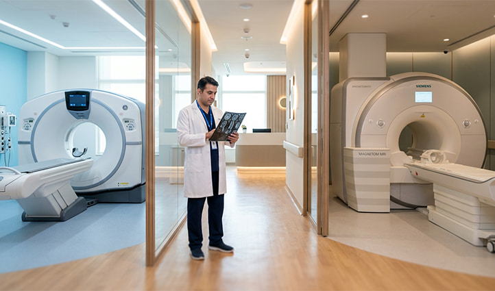
The Human Element of Diagnostics
It is easy to focus entirely on the machines, but a scanner is only as good as the person interpreting the images. A sophisticated MRI can produce thousands of high-definition slices, but it takes a highly trained eye to spot a microscopic lesion hidden among healthy tissue.
This is exactly why choosing where you have your scans done matters so much. Attending a dedicated radiology hospital in Dubai ensures you are scanned on the latest-generation equipment, which translates to faster scans, lower CT radiation doses, and vastly superior image resolution.
More importantly, it guarantees that your scans are being reviewed by a subspecialist. Having an experienced radiologist in Dubai read your images means you receive a precise, nuanced report. They do not just look for obvious abnormalities; they correlate the imaging with your specific medical history to give your treating doctor the exact information needed to chart the right course of treatment.
Frequently Asked Questions
Will I feel anything during a CT or MRI scan?
No. Both CT and MRI scans are entirely painless. You will not feel the magnetic fields of the MRI or the X-rays of the CT. The only discomfort might be lying still for an extended period, or a slight warmth if an IV contrast dye is used.
Why do I sometimes have a CT and an MRI on the same day?
Because they show different things, a doctor might order a CT scan to quickly rule out a bone fracture or internal bleeding, and immediately follow it with an MRI to get a detailed look at the surrounding soft tissue or spinal cord.
Is the radioactive tracer in a PET scan dangerous?
It's not dangerous. The amount used is small, clears the body quickly, and the dose is worked out specifically to make sure the scan is useful without unnecessary exposure. It's one of the more routine parts of the procedure.